Friday 30 March 2012

Prevalence and Incidence

Prevalence and Incidence of Adrenoleukodystrophy 

Prevalence is a frequently used epidemiological measure of how commonly a disease or condition occurs in a population. Prevalence measures how much of some disease or condition there is in a population at a particular point in time. The prevalence is calculated by dividing the number of persons with the disease or condition at a particular time point by the number of individuals examined. 


The incidence of a disease is another epidemiological measure. Incidence measures the rate of occurrence of new cases of a disease or condition. Incidence is calculated as the number of new cases of a disease or condition in a specified time period (usually a year) divided by the size of the population under consideration who are initially disease free. 


It has been found that the prevalence of adrenoleukodystrophy or the number of individuals with this disease is 1 in every 200,000.


The incidence was approximated to be 1 in every 100,000.


This disease has also been found in all races and on all of the continents. 


Keep in mind that these estimates are referring to all the forms of adrenoleukodystrophy, the child and adult forms. 


References


http://www.rightdiagnosis.com/a/adrenoleukodystrophy/prevalence.htm 


http://ghr.nlm.nih.gov/condition/x-linked-adrenoleukodystrophy


Alisha M.

Wednesday 28 March 2012

Stages

Classic childhood form (X-linked adrenoleukodystrophy)

In this case of ALD, the child usually develops normally usually up to 3 or 4 years of age. The first noticeable problems are behavioral changes such as hyperactivity, difficulty paying attention, aggression, poor academic performance, difficulty reading and comprehending and abnormal withdrawal. In this stage, children are most often misdiagnosed with ADHD. As the disease progresses, more serious difficulties occur such as loss of hearing and vision. The child is unable to discriminate sounds and has difficulty understanding speech. Also, they may develop an inability to see visual information on the left or right side, may not be able to see clearly, and double vision. This eventually leads to deafness and blindness. ALD patients will then experience difficulty walking, swallowing, have learning impairments such as poor memory, difficulty in speech articulation due to the inability to control muscles involved in speech, fatigue and seizures. About 33% of patients with ALD develop seizures, which may be the first sign of ALD. 90% of children develop insufficient production of hormones by adrenal glands by the time signs of the disease are first noticed. From the onset of the initial symptoms, patients can be in a vegetative state within 6 months to 2 years, although rate of progression varies in different patients. Death can occur anytime after symptoms begin, but usually occur within 1 to 10 years.

Adult-onset form (adrenomyeloneuropathy)

This form of ALD usually occurs between the ages of 21 and 35. It progresses more slowly than the childhood form, with the first symptoms being stiffness and weakness in legs that worsen over time. Patients develop ataxia (impairment in coordinating movements), difficulty walking, sensory loss, and loss of bladder control (no sphincter control). About half of patients show brain damage, with 33% having myelin loss, and 10-20% showing severe brain damage. This usually results in total disability and death. 70% have insufficient production of hormones by the adrenal glands. This type of ALD usually progresses over a 5 to 15 year period, but can go progressing for decades.

Nevena V.

References:


Moser MW, Mahmood A, Raymond GV (2007) X-liked adrenoleukodystrophy. Nature Review 3(3): 140-151.

Tuesday 27 March 2012

Hematopoietic Stem Cell Gene Therapy with a Lentiviral Vector

As discussed beforehand, ALD is a disease deficient in the ALD protein, an adenosine triphosphate-binding cassette transporter encoded by the ABCD1 gene involved in fatty acid degradation.  Researchers have found a novel way to replace this dysfunctional gene with the wild-type by using meas of gene therapy. Many of the therapies for ALD today involve bone marrow transplantation. The long-term benefits of bone marrow transplantation are mediated by the replacement of brain microglial cells derived from donor bone marrow myelo-monocytic cells. Unfortunately, bone marrow transplantation carries a high risk of mortality and it can be expecially difficult to find a matched donor. Researchers introducing gene therapy has reasoned that hematopoietic stem cell gene therapy can be a more appropriate therapeutic alternative.

In this therapy, CD34+ cells were removed from two patients and a lentiviral vector encoding the wild-type ABCD1 gene was infused into the hematopoietic cell and then reinfused back into the patients. An HIV-derived vector was used to deliver the therapeutic gene into patients cells.


After 24 to 30 months, there was a detected polyclonal reconsitution with 9 to 14% of granulocytes, monocytes, and T and B lymphocytes expressing the ALD protein.

In the graphs protrayed above, there is an increased percentage of lymphocytes/monocytes, and CD+ cells expressing the ALD protein. There is also a decreased concentration of C26:0/C22:0 fatty acids after gene therapy.

14 to 16 months after infusion, MRI scans show progressive demyelination stopped as depicted below.


Nevena V

References:

http://www.sciencedaily.com/releases/2009/11/091105143706.htm

Cartier N. et al. (2009) Hematopoietic Stem Cell Gene Therapy with a Lentiviral Vector in X-linked Adrenoleukodystrophy. Science 326: 818-823.

Monday 26 March 2012

Treatment with Lorenzo's Oil

Lorenzo’s Oil is a 4:1 mixture of glyceryl trioleate (oleic acid - monounsaturated fatty acid) and glyceryl trierucate (erucic acid - monounsaturated fatty acid). It has been found that oral administration of this oil in cerebral ALD patients is able to successfully normalize levels of VLCFA in the plasma, only for boys that are largely asymptomatic. There therapy was first introduced in 1981, with intentions to reduce the level of very long chain fatty acids (VLCFA) in the plasma. It was found that a diet alone with reduced intake of saturated VLCFA did not alter plasma C26:0 levels, due to endogenous synthesis of these fatty acids. In 1986, the addition of monounsaturated oleic acid reduced the levels and the rate of biosynthesis of saturated VLCFA in cultured skin fibroblasts of patients with ALD. From this, it was found that oral administration of gylceryl trioleate for a period of 3 to 4 months significantly lowered plasma C26:0 levels by approximately 50%. Furthermore, in 1989, Augusto Odone, the founder of Lorenzo’s Oil, added erucic acid to the oil, on the basis of a review of lipid manipulation in animal studies and reports that erucic acid and saturated long-chain fatty acids are elongated by the same microsomal enzyme system. The unsaturated fatty acids in Lorenzo’s Oil competitively compete for chain elongation with saturated fatty acids resulting in reduced endogenous synthesis of VLCFA. This led to the final component in Lorenzo’s Oil, and has been shown to normalize levels of saturated VLCFA and delay the progression of neurologic abnormalities within 4 weeks in most patients.

Lorenzo’s Oil is still popularly used in many ALD patients, but has received inconclusive results within studies. A study conducted by Moser et al. (2005) followed-up 89 asymptomatic boys with X-ALD between the years of 1989 and 2002 that have been treated with Lorenzo’s Oil. The oil was taken orally in a dosage that provided 20% of caloric intake while other fat intake was only limited to 10% to 15% of total calories. Patients were followed up at 6-month intervals. 74% (66) of studied patients were well upon their last follow-up, meaning that they exhibited normal neurological status and normal brain MRI results. Results from the study are shown in the table below after the final follow-up. From this study, it is recommended that Lorenzo’s Oil therapy be offered to male patients with ALD who are neurologically asymptomatic, have normal brain MRI results and are at risk of developing cerebral ALD. Intensive Lorenzo’Oil therapy during the ages at which the risk for cerebral ALD is greatest (boys younger than 7 years) may protect against this phenotype until patients reach ages at which the risk for cerebral ALD diminishes (after 10 years of age).


The administration of Lorenzo’s Oil has only been successful in halting the progression of the disease in patients that are largely asymptomatic, and has limited success in patients that already have neurological abnormalities and are symptomatic. There is evidence that dietary therapy can reduce the levels of VLCFA in the plasma, adipose tissue and liver but no significant reduction is observed in the brain (possibly due to the fact that erucic acid cannot enter the brain at a significant quantity). There is also no evidence of a clinically relevant benefit from dietary treatment with oleic and erucic acids in patients with adrenomyeloneuropathy. Early detection of this disease is crucial in order to obtain the benefits of the oil.

Nevena V.

References:


Moser H, Dubey P, Fatemi A (2004) Progress in X-linked adrenoleukodystrophy. Current opinion in neurology 17(3): 263-269.

Moser H et al. (2005) Follow-up of 89 Asymptomatic Patients With Adrenoleukodystrophy Treated With Lorenzo’s Oil. Archives of Neurology 62: 1073-1080.

Aubourg et al. (1993) A two-year trial of oleic and erucic acids ("Lorenzo's Oil") as treatment for adrenomyeloneuropathy. The New England Journal of Medicine 329(11): 745-752.

Rasmussen M., Moser A., Borel J., Khangoora S., Moser H. (1994) Brain, liver, and adipose tissue erucic and very long chain fatty acid levels in adrenoleukodystrophy patients treated with glyceryl trierucate and trioleate oils (Lorenzo's Oil). Neurochemical Research 19(8): 1073-1082.


Wednesday 21 March 2012

Lovastatin as a Treatment for Adrenoleukodystrophy


Lovastatin for X-Linked Adrenoleukodystrophy

 Lovastatin

In a study conducted by Singh et. al they have shown in animal studies that lovastatin can be used as a therapy for adrenoleukodystrophy. Lovastatin is a 3-hydroxy-3-methylglutaryl coenzyme A reductase inhibitor and sodium phenylacetate, which inhibits mevalonate pyrophosphate decarboxylase. This then inhibits the induction of inducible nitric oxide synthase and proinflammatory cytokines involved in the pathogenesis of neurological damage in X-linked adrenoleukodystrophy. For those not in a science major here is some definitions that will help:

-       A 3-hydroxy-3-methylglutaryl coenzyme A reductase inhibitor is the rate-controlling enzyme of the mevalonate pathway, the metabolic pathway that produce cholesterol
-         Sodium phenylacetate is an aromatic fatty acid, which displays cell growth inhibition, malignant phenotype reduction and cell differentiation
-       A cytokine is a are small cell signaling molecules that are usually produced during an immune reaction

Lovastatin has been shown to increase intracellular cyclic AMP and protein kinase A activity and normalize the levels of very-long chain fatty acids in skin fibroblasts from patients with childhood adrenoleukodystrophy.

The study was conducted by treating patients with 20 mg of lovastatin per day for two weeks, if not side effects occurred the dose was increased to 40 mg a day.  The fatty acids were measured throughout the study to observe the effects. According to their results the plasma levels of very-long chain fatty acids declines in each patient that completed the trial after 6 months.  These results suggested that lovastatin treatment may reduce the very-long chain fatty acids in adults with adrenoleukodystrophy, without any significant side-effects.  The effects of lovastatin are believed to be due to the drug blocking the induction of inflammatory mediators of neurological damage in adrenoleukodystrophy.

A comparative study conducted by Engelen et. al have stated that lovastatin should not be used for a treatment of adrenoleukodystrophy. The conducted their own study where they gave patient with adrenoleukodystrophy a 40 mg dose of lovastatin once daily compared to a placebo dose. The trial was designed to investigate whether lovastatin has a biochemical effect in vivo in patients with X-ALD. They concluded that lovastatin leads to a small decrease in levels of C24:0 and C26:0 in plasma. They also took into account that VLCFAs (very-long-chain fatty acids) are water soluble and therefore only a small amount binds to the albumin in the blood and most bind to the LDL cholesterol.  LDL did not decrease with the lovastatin dose and therefore they considered the results to be nonspecific due to the fact that LDL did not decrease.
With the opposing results of these two studies it is clear that more research is needed to study the effects of lovastatin in patients with adrenoleukodystrophy.  Whether this drug can be used as a therapy is still unclear and more time and resources must be placed on the study of this drug to determine if it holds any positive benefits.

References

Singh, I., Khan, M., Key, L. & Pai, S. 1998, Lovastatin for X-Linked Adrenoleukodystrophy. The New England Journal of Medicine, 339: 702-703

Engelen, M., Ofman, R., Dijkraaf, M., Hijzen, M. & Wardt, L. Lovastatin in X-linked Adrenoleukodystrophy. 2010. The New England Journal of Medicine, 362, 276-277.

Alisha M.

Wednesday 14 March 2012

Bone Marrow Transplant Therapy

Adrenoleukodystrophy: Bone Marrow Transplant Therapy

Bone marrow transplants have been used with the intention to treat storage diseases such as adrenoleukodystrophy The experiments that have been conducted has been shown to increase the life expectancy of patients that received the transplant compared to patients that did not.

Bone marrow transplantation works by the engraftment from normal donors providing cells that express a normal ratio of oxidation of C224:0 compared to C16:0. This graft has been shown to persist for as long as 8 years. The rationale for BMT in X-ALD has relied on the hypothesis that functional bone-marrow cells from the donor could cross the blood-brain-barrier in the recipient and exert favorable effects on the mechanisms leading to demyelination. Subsequently, short-term benefits of BMT in childhood cerebral X-ALD were reported.
This graph demonstrates the post-BMT effects in four patients with adrenoleukodystrophy. The shaded regions correspond to abnormal C24/16 ratio. In each graph it shows that after a bone marrow transplant the recipient show a normal C24/16 ratio.

Other neuropsychological evaluations have shown that patients destine to develop the severe form of childhood onset of cerebral adrenoleukodystrophy who receive a bone marrow transplant show evidence of stability and/or improvement, especially in verbal memory.

MRI observations have also indicated an abatement of the degenerative processes in the brain compared to transplanted patients. In the experiment done by Shapiro et. al, 2000, 12 patients with childhood onset of adrenoleukodystrophy were followed for 5-10 years after a bone marrow transplant. MRI showed complete reversal of abnormalities in two patients and improvement in one. One showed no change and all 8 patients who showed an initial period of continued demyelination stabilized and remained unchanged thereafter. Motor function remained normal or improved after BMT in 10 patients. Verbal intelligence remained with in the normal range for 11 patients. Plasma VLCFA concentrations decreased by 55% and remained slightly above the upper limit of normal.

This study of twelve transplanted and grafted cerebral X- ALD patients confirms the long-term beneficial effect of BMT when done early in the disease course.
Krivit et. al (1995) performed an experiment with 5 sets of brother in the same family who both have a likelihood of developing childhood adrenoleukodystrophy. The younger brothers of each pair were given the bone marrow transplant. In each, the non-transplanted patient has dies and the transplanted patient remained alive.


Presymptomatic patients are now being treated via two methods:
1) Bone marrow transplantation
2) Dietary manipulation as well as GTE:GTO supplementation to provide normalization of plasma VLCFA. There is hope that presymptomatic individuals who are on the diet may inhibit the onset of neurological deterioration

Things to consider before a bone marrow transplant:
  • Correction of increased VLCFA is recommended before transplantation. Cell membrane are abnormal when VLCFA are elevated, red blood cells are an example. In high plasma VLCFA erythrocytes have higher mechanical fragility. When VLCFA in the plasma is within the normal reference range this fragility disappears.
  • When C26:0 is below 0.4 ug/ml there has been minimal difficulty during the transplantation. If these levels are increased however difficulties have occurred. To solve this problem artificial lipid emulsions should be excluded.
  • Platelet number ad function is also disrupted during dietary restriction and use of Lorenzo’s oil. The diet should be discontinued when transplantation is begun as well as addition of mandatory platelet transfusion.
  • Severe and obvious neurological disabilities have been showed to lead to increased deterioration with bone marrow transplants. Eligibility should be carefully evaluated
  • Graft rejection has also caused disadvantages to this procedure. The removal of T cells by elutriation will increase the availability of donors for those without HLA sibling or unrelated phenotypic identical ones.

After the transplantation it has been prescribed that no dietary restrictions need to be taken. No correlation of IQ with VLCFA subsequent to bone marrow transplantation has been found. Post transplantation data has indicated that all of the patients have elevated levels of VLCA despite full engraftment as measured by donor DNA and normal levels of oxidation activity of white cells.
Future goals for this therapy is to increase the use of elutriation of non-genotypic marrow and of cord-blood hematopoietic stem may vastly increase sources for donor cells, which will allow greater freedom from therapeutic decision making.

References

Krivit, W., Lockmam, A., Watkins, P., Hirsch, J. & Shapiro O. 1995. The future for 
treatment by bone marrow transplantation for adrenoleukodystrophy, metachromatic leukodystrophy, globoid cell leukodystrophy and Hurler syndrome. The Jornal of Inherited Metabolic Diseases, 18; 398-412.  

Shapiro, E., Krivit, W., Lockman, L., Lambaque, I., Peters, C., Cowan, M., Harris, R., Blanche, S., Bordigoni, S., Loes, D., Ziegler, R., Crittenden, M., Ris, B., Cox, C., Moser, H., Fischer, A. & Aubourg, P. 2000. Long-term effect of bone-marrow transplantation for childhood-onset cerebral X-linked adrenoleukodystrophy. The Lancet, 356; 713 - 719.

Alisha M.

Saturday 10 March 2012

Diagnostics

After the appearance of potential ALD-like symptoms (ataxia, spasticity, deafness, visual deficits, behavioral disturbances), patients are encouraged to undergo diagnostic testing. Neuroimaging techniques such as Magnetic Resonance Imaging (MRI), Computed Tomographic (CT) scans, and Positron Emission Tomography (PET) scans are valid techniques for examining cerebral demyelination in patients with X-linked ALD and usually provide the first lead to diagnosis in relation to symptoms.

The following images are from a 3 year case report of a 24-year old male diagnosed with ALD with a 10 year history (Volkow et al. 1987). Imaging was first conducted in 1984 where the patient had progressive loss of visual acuity. Ten months later (1985), the patient returned and neurological examination revealed progression of visual defect and decreased proprioception. 8 months later, the patient experienced acute psychosis, delusions, almost complete visual loss, hallucinations, ataxia, and progressive loss of memory and attention span.

This first image depicts a CT scan of the patient with decreased density in white matter (shown by decreased contrast) over the three year case study.

 These images show an MRI scan of the patient in 1985, and then in 1986. There is further edema (excess swelling) into white matter tracts (right image) and demyelination.

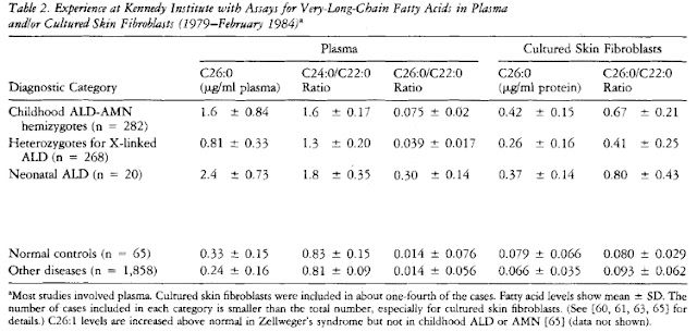
After clear observation of brain damage and demyelination, a plasma, fibroblast, or red blood cell assay is the most widely used and convenient step for a definitive diagnosis. Measurements of very long chain fatty acids levels (VLCFA), mainly the fatty acid hexacosanoate (C26:0), in red blood cell phospholipids or total plasma lipids are taken. Elevated C26:0 levels, as wells as elevated C24:0/C22:0 and C26:0/C22:0 ratios indicate a peroxisomal disorder in which these fatty acids are unable to undergo oxidation and may indicate ALD. A fibroblast assay may also take place to detect elevated VLCFA levels. The table below shows increased C26:0 levels and ratios in Childhood Adrenoleukodystropy-Adrenomyeloneuropathy (ALD-AMN), Heterozygotes for X-linked ALD (mothers - carriers), and neonatal ALD (Moser et al. 1984):


Additionally, a family history of the disease is suggested. It is recommended that mothers (the carriers) also have their plasma and fibroblast levels of hexacosonoate examined for a more definitive diagnosis of X-ALD in their male offspring. It has been observed that female heterozygotes also exhibit elevated C26:0 levels and ratios and may potentially exhibit some symptoms of ALD. Furthermore, due to increased chance of false negatives in plasma and fibroblast measurements, a mutational analysis is recommended for all potential carriers. The table below shows elevated C26:0 levels in diagnosed heterozygotes for X-linked ALD (Moser et al. 1984):


Currently, prenatal diagnosis is available in where amniocytes are obtained by amniocentesis and C26:0 measurements are taken from cultured cells. Abnormal results could result in abortion of the fetus.

Flowchart of recommended actions of diagnosis (Shimozawa 2011):


Nevena V.

References

Moser HW, Mahmood A, Raymond GV (2007) X-linked adrenoleukodystrophy. Nature:Clinical Practice. 3(3):140-151.

Moser HW, Moser AE, Singh I, O'Neill BP (1984) Adrenoleukodystrophy: Survey of 303 Cases: Biochemistry, Diagnosis, and Therapy. Ann Neurol. 16: 628-641.

Shimozawa N (2011) Molecular and clinical findings and diagnostic flowchart of peroxisomal diseases. Brain and Development. 33: 770-776.

van Geel Bjorn, Assies J, Wanders RJA, Barth PG (1997) X linked adrenoleukodystrophy: clinical presentation, diagnosis, and therapy. Journal of Neurology, Neurosurgery, and Psychiatry. 63: 4-14.

Volkow ND, Patchell L, Kulkarni MV, Reed K, Simmons M (1987) Adrenoleukodystrophy: Imaging with CT, MRI, PET. The Journal of Nuclear Medicine. 28(4): 524-527.

Main Forms and Symptoms

Categories
Adrenoleukodystrophy presents in three main categories:

  • Childhood cerebral form -- which appears in mid-childhood (at ages 4 - 8)
    • affects white matter and related cells in the brain
  • Adrenomyeloneuropathy -- occurs in men in their 20s or later in life
    • affects axonal tracts of the spinal cord
  • Impaired adrenal gland function (called Addison disease or Addison-like phenotype) -- adrenal gland does not produce enough steroid hormones
These categories each present symptoms.
Symptoms 
Childhood cerebral type:
  • Changes in muscle tone, especially muscle spasms and spasticity
  • Crossed eyes (strabismus)
  • Decreased understanding of verbal communication (aphasia)
  • Deterioration of handwriting
  • Difficulty at school
  • Difficulty understanding spoken material
  • Hearing loss
  • Hyperactivity
  • Worsening nervous system deterioration
    • Coma
    • Decreased fine motor control
    • Paralysis
  • Seizures
  • Swallowing difficulties
  • Visual impairment or blindness
Adrenomyelopathy:
  • Difficulty controlling urination
  • Possible worsening muscle weakness or leg stiffness
  • Problems with thinking speed and visual memory
Adrenal gland failure (Addison type):
  • Coma
  • Decreased appetite
  • Increased skin color (pigmentation)
  • Loss of weight, muscle mass (wasting)
  • Muscle weakness
  • Vomiting


Alisha M.
References from: http://www.ncbi.nlm.nih.gov/pubmedhealth/PMH0002165/

Thursday 1 March 2012

About Adrenoleukodystrophy

Adrenoleukodystrophy is an X-linked peroxisomal disorder that affects the white matter of the central nervous system, adrenal cortex and testes in boys. The incidence is rare, in that only 1:50,000 males are affected with the disease. People diagnosed with X-linked ALD have elevated plasma levels of saturated very long chain fatty acids (VLCFA – C22 or longer), and for reasons currently not clearly understood, these elevated  levels of VLCFA are toxic to the body, primarily the myelin sheath in the central nervous system. VLCFA are broken down in peroxisomes rather than, like most fatty acids, in the mitochondria due to their large size.
Research has indicated that a gene defect located in Xq28 on the X chromosome is the primary cause of this disease. Xq28 encodes for peroxisomal membrane protein, ALD-P (Adrenoleukodystrophy Protein). ALD-P belongs to the adenosine triphosphate-binding cassette (ABC) superfamily of transmembrane transporters. ABC proteins transport a variety of molecules ranging from ions to proteins across extra- and intra-cellular membranes. An increasing number of mutations (mainly missense) in the ABCD1 gene results in the absence or defective ALD-P, which is responsible for the transfer of VLCFA to peroxisomes for β-oxidation. Furthermore, the metabolism of these fatty acids are activated by their coenzyme A thioesters, acyl-CoA synthetase. Coenzyme activity has also been found to be impaired in ALD patients. Activation of VLCFA by very long chain acyl-CoA synthetase (VLCS) takes place in the peroxisome organelle. Lack of VLCS prevents this organelle from catabolizing VLCFA via β-oxidation.

Due to the inability of VLCFA to be oxidized, an autoimmune or cytokine-mediated inflammatory response normally occurs, leading to cerebral white matter demyelination and scarring due to gliosis.

Nevena V.
References

Kemp S, Pujol A, Waterham HR, van Geel BM, Boehm CD, Raymond GV, Cutting GR, Wanders RJA, Moser HW (2001) ABCD1 Mutations and the X-linked Adrenoleukodystrophy Mutation Database: Role in Diagnosis and Clinical Correlations. Human Mutation. 18(6): 499-515.

Melhem ER, Barker PB, Raymond GV, Moser HW (1999) X-Linked Adrenoleukodystrophy in Children: Review of Genetic, Clinical, and MR Imaging Characteristics. American Journal of Roentgenology. 173(6): 1575-1581.

http://www.x-ald.nl/